Trade Schools, Colleges and Universities
Join Over 1.5 Million People We've Introduced to Awesome Schools Since 2001

Trade Schools, Colleges and Universities
Join Over 1.5 Million People We've Introduced to Awesome Schools Since 2001
Independent dental radiography study resource
Practice the decision, not just the definition. Take an instant quiz, try a 60-minute mock test, or troubleshoot digital dental X-ray artifacts visually. No sign-up.
By Chris Gaglardi · Trade-Schools.net · Prepared October 9, 2026 · Educational material
The interactive practice workspace is loading. You can still use the evidence-based study guide below if scripting is unavailable.
The Radiation Health and Safety (RHS) exam covers dental imaging techniques, radiation protection, and infection prevention. According to DANB, the examination has 75 multiple-choice questions and a 60-minute testing time. DANB uses computer-adaptive testing (CAT): the questions change based on performance, and examinees cannot return to earlier items. Check the official DANB RHS page before applying.
Reported DANB exam results use a 100–900 scaled score, with a passing score of 400. That is not the same as getting 40%, 50%, or any other fixed percentage of questions right. Because this site's practice test is a fixed, uncalibrated question set, it reports raw practice accuracy only. We will not invent a passing probability from it. DANB explains the scoring method.
| RHS content area | DANB weight | Our practice questions |
|---|---|---|
| Purpose and Technique | 50% | 37 |
| Radiation Characteristics and Protection | 25% | 19 |
| Infection Prevention and Control | 25% | 19 |
Study digital radiography. DANB removed traditional film-processing topics from RHS testing in July 2022. The full outline is broader than the questions on this page: it also covers anatomy, image mounting, patient management, legal image-handling duties, radiation biology, and infection prevention. Receptor types, positioning, image quality, patient and operator protection, and infection control remain relevant. Check DANB's RHS content outline (PDF) and the latest application documents, not an undated exam-dump site.
Certain positioning and processing errors share visual features, so context matters as much as the apparent pattern. The visual lab above lets you practice making two decisions: identify the likely cause, then choose an appropriate correction. The schematic illustrations isolate common technique concepts; they are not diagnostic radiographs or a substitute for interpreting actual clinical images.
Technique corrections should be taught and supervised by qualified clinicians. For example, a distorted image could have several contributing causes; the training case isolates one variable to make the physics easier to learn.
The American Dental Association's February 2024 recommendations no longer advise routine lead aprons or thyroid collars for patients during dental imaging. Shielding can obscure anatomy and force a repeat exposure. Appropriate patient selection, digital receptors, precise beam collimation, and sound positioning are better-supported tools for limiting unnecessary radiation.
The ADA and AAOMR's January 2026 patient-selection recommendations emphasize taking images when clinically indicated for the individual patient, rather than automatically on a fixed schedule. A dentist, not an independently acting assistant, determines the clinical need for an imaging examination.
Radiation safety also includes reducing preventable retakes, using authorized imaging protocols, remaining behind designated operator shielding, and never using your hand as a replacement for a proper receptor holder. Always follow applicable regulations, clinician supervision, and the equipment manufacturer's instructions.
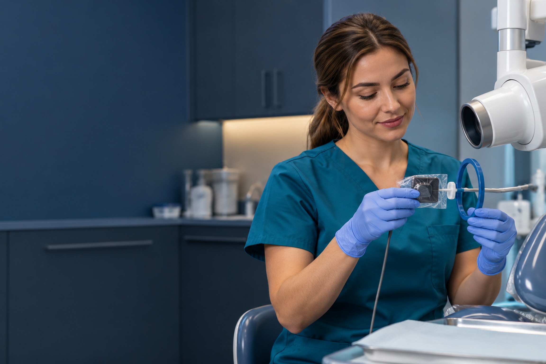
Intraoral digital sensors contact oral mucous membranes, so the CDC classifies them as semicritical devices. Use an FDA-cleared barrier to reduce contamination, then carry out the device's validated reprocessing process between patients.
If a digital sensor can tolerate heat sterilization or high-level disinfection, CDC recommends those methods after cleaning. When it cannot tolerate them, CDC advises an FDA-cleared barrier plus cleaning and an EPA-registered hospital disinfectant with intermediate-level activity between patients. Manufacturers specify which methods are compatible. Neither “all sensors are safe after a quick wipe” nor “all digital sensors must be autoclaved” is reliable advice.
Reusable, heat-tolerant intraoral holders and bite blocks need appropriate cleaning and sterilization. Wear suitable PPE for contact and anticipated splash or spray, perform hand hygiene, change disposable barriers between patients, and keep contaminated sleeves away from clean scanning and computer areas. Review the CDC's dental standard precautions.
Start with the 15-question diagnostic. If one domain is weak, revisit that section of DANB's current outline and work through your course materials. Then use the illustrated lab to practice error recognition. Save the 75-question timed set for a separate session when you can concentrate for an hour.
Do not interpret one low practice percentage as proof you need paid school. It could reflect unfamiliar terminology, a narrowly missed technique concept, or a knowledge gap best addressed by an instructor and targeted review. Likewise, a high score here does not guarantee a pass on a computer-adaptive examination.
Learn how dental assistants enter the field. Compare your state requirements before paying for any program. If training makes sense, investigate dental assistant programs and schools. Those school decisions are separate from reviewing an RHS exam score.
Need a refresher on orientation and numbered teeth? Work through our free tooth-numbering practice tool. Comparing assistant versus hygienist duties? See our side-by-side career guide.
DANB lists 75 multiple-choice questions and 60 minutes. The official exam uses computer-adaptive testing; our 75-question practice uses a fixed bank.
DANB's scaled passing standard is 400 on a 100–900 scale. There is no valid conversion from an ordinary practice percentage to a DANB score or pass probability.
DANB states that the standalone RHS exam has no eligibility prerequisites. Permission to take dental X-rays on patients depends separately on state requirements and an authorized scope of duties.
No. RHS is only one component used in DANB's CDA and NELDA credential pathways. Review complete credential eligibility and examination requirements with DANB.
No. Laws governing radiographic duties and state registration vary. Consult your dental board and DANB's state directory before undertaking clinical duties.
No. The ADA's 2024 guidance no longer recommends routine patient shielding, but state or local rules may still apply. Follow applicable law and the supervising dentist's protocol.
Not as a universal rule. CDC recommends an FDA-cleared barrier plus device-compatible cleaning and sterilization or disinfection. For sensors unable to tolerate heat sterilization or high-level disinfection, use a barrier plus cleaning and EPA intermediate-level disinfection, according to manufacturer instructions.